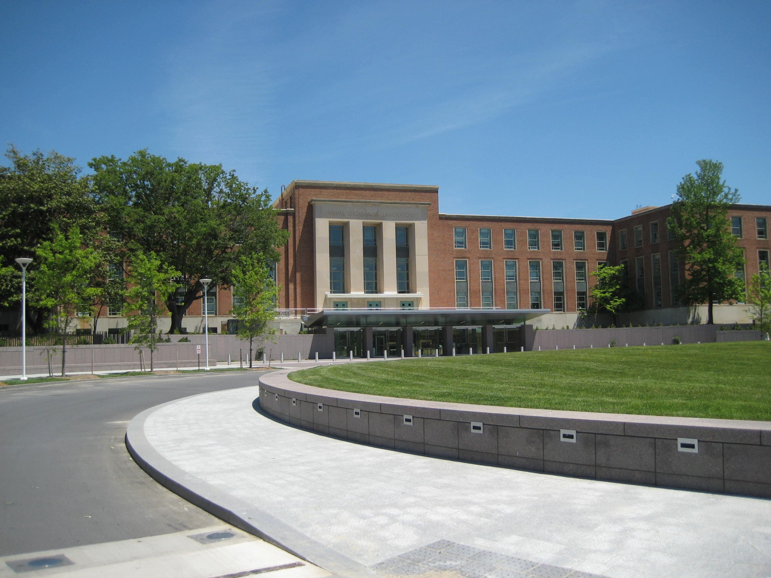
Three Scientists Share the 2026 Nobel Prize in Medicine for Teaching Brain Cells to Answer to Light
The Nobel Assembly at Karolinska Institutet has awarded the 2026 Nobel Prize in Physiology or Medicine to Karl Deisseroth, Peter Hegemann and Georg Nagel for the discovery of light-gated ion channels in a single-celled alga and their transformation into optogenetics, a method for switching nerve cells on and off with light.
Just News Desk, Correspondent||6 min read
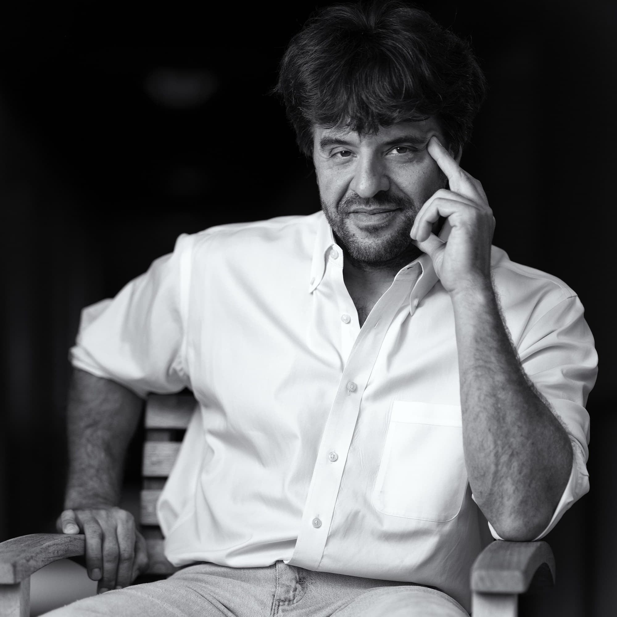
STOCKHOLM - Three scientists whose work began with a simple question about how a green alga finds the light have been awarded the 2026 Nobel Prize in Physiology or Medicine, the Nobel Assembly at Karolinska Institutet announced on 5 October. Karl Deisseroth of the Howard Hughes Medical Institute and Stanford University, Peter Hegemann of Humboldt University of Berlin and Georg Nagel of the University of Würzburg share the prize "for their discoveries concerning light-gated ion channels and optogenetics."
The method the three made possible, known as optogenetics, allows researchers to switch specific nerve cells on with a pulse of light and to watch what follows in a living brain. In its announcement, the assembly said the laureates "have laid the foundation of a new era in neuroscience."
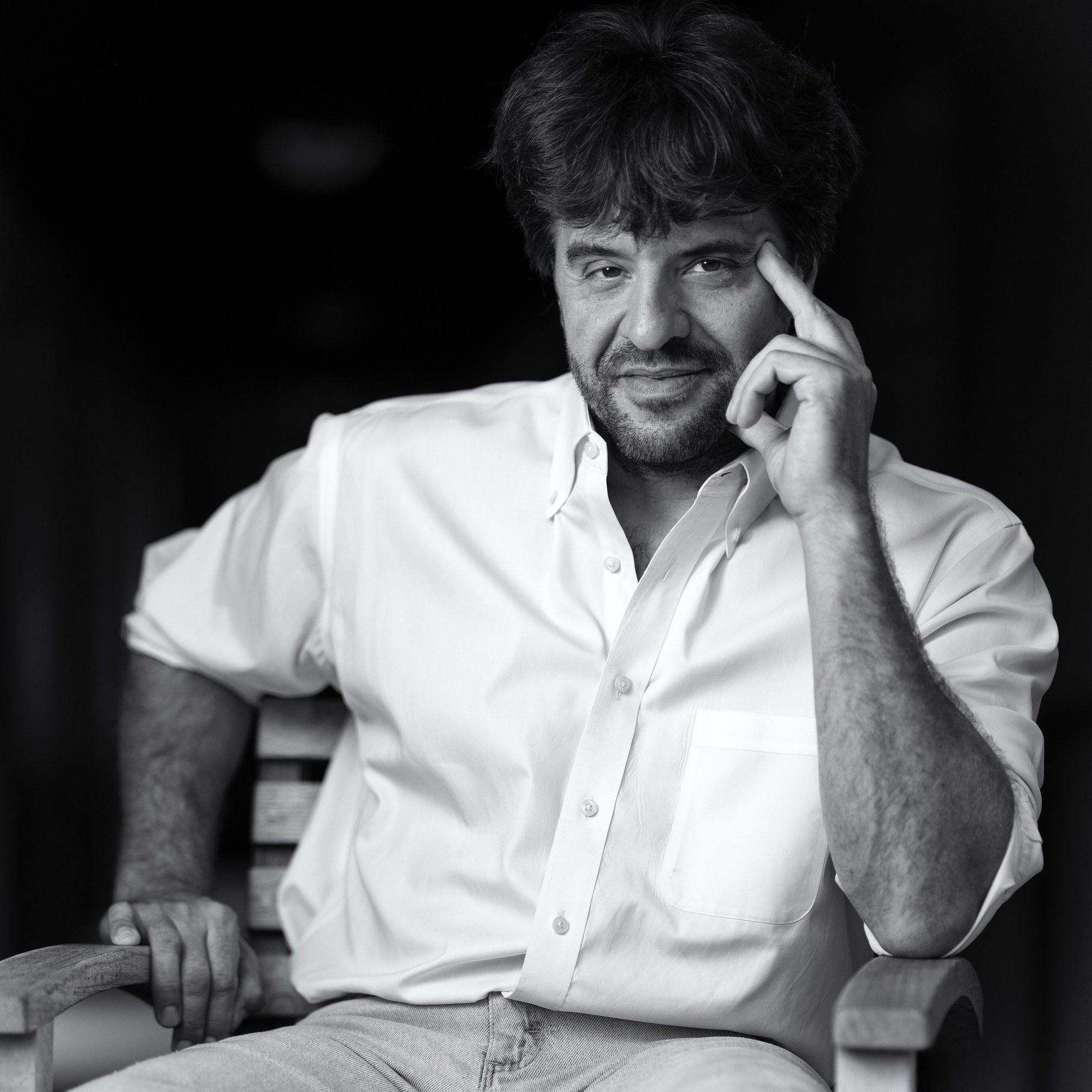
Karl Deisseroth, a professor of bioengineering and of psychiatry and behavioral sciences at Stanford University and a Howard Hughes Medical Institute investigator, photographed on 20 September 2022. Photo: Christopher Michel, own work, via Wikimedia Commons (CC BY-SA 4.0). Source page: https://commons.wikimedia.org/wiki/File:Karl_Deisseroth_by_Christopher_Michel_01.jpg
For much of the 20th century, the assembly noted, scientists could link regions of the brain to particular functions but could not prove which cells caused what. The picture they assembled was "like a sketch map, full of question marks and unknowns."
The path out of that uncertainty began with Dr. Hegemann's curiosity about Chlamydomonas, a single-celled alga that swims toward light. In the early 2000s, according to the assembly, he and Dr. Nagel discovered channelrhodopsin, a protein on the surface of the cell. When blue light strikes it, a channel opens through the protein, charged ions flow into the cell and an electrical impulse results. The two found that whatever cell they placed the protein in became sensitive to light. The work was done at Max Planck institutes in Germany: Dr. Hegemann's at the Max Planck Institute for Biochemistry in Martinsried, and Dr. Nagel's at the Max Planck Institute for Biophysics in Frankfurt.
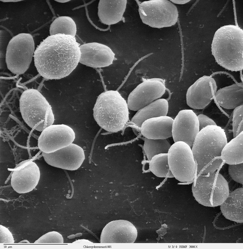
Chlamydomonas reinhardtii, the single-celled green alga in which channelrhodopsin was found, seen under a scanning electron microscope. Image: Dartmouth Electron Microscope Facility, Dartmouth College, via Wikimedia Commons (public domain). Source page: https://commons.wikimedia.org/wiki/File:Chlamydomonas6-1.jpg
Dr. Deisseroth carried the discovery into the nervous system. He introduced the gene for channelrhodopsin into nerve cells taken from rats and, by shining blue light on them, triggered a nerve signal, a result he published in 2005. Two years later, the assembly said, he made the light-controlled switch work in the brains of living mice.
"Optogenetics provides opportunities for mapping the brain in a way that we could once only dream of," Per Svenningsson, chair of the Nobel Committee for Physiology or Medicine, said in the announcement.
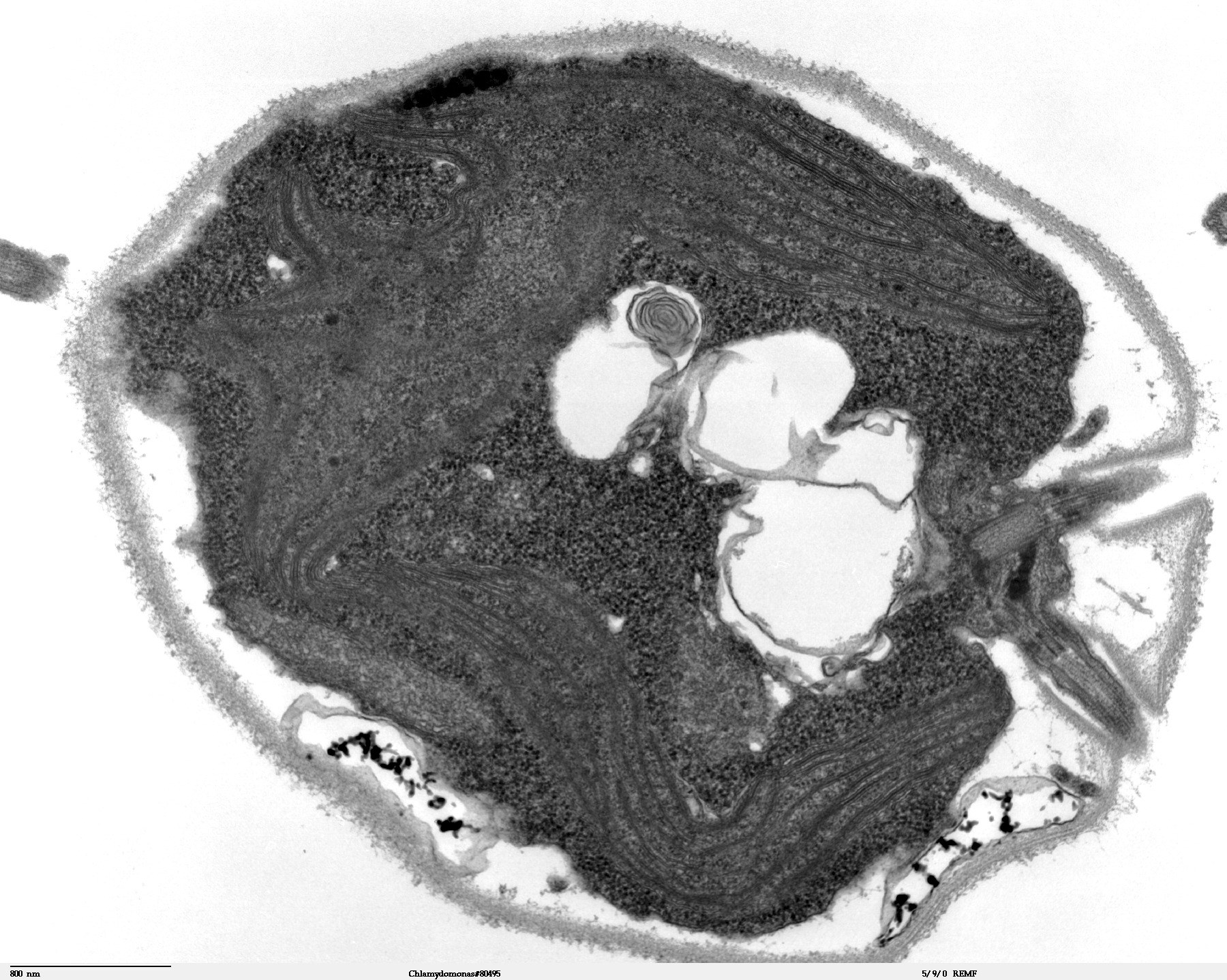
A thin section through a single Chlamydomonas cell under a transmission electron microscope, showing the chloroplast, vacuoles, eyespot, flagellar basal body area and cell wall. Image: Dartmouth Electron Microscope Facility, Dartmouth College, via Wikimedia Commons (public domain). Source page: https://commons.wikimedia.org/wiki/File:Chlamydomonas_TEM_01.jpg
The method has spread widely. Using it, researchers have revealed neural circuits that govern specific memories, feelings and behaviors relevant to neurological and psychiatric disorders, according to the assembly, and in clinical medicine researchers are using it in attempts to restore sight to people with visual impairment.
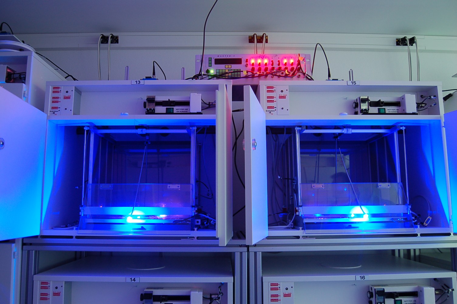
Laboratory cages for rats fitted with LED commutators that allow researchers to study animal behavior during optogenetic stimulation, photographed 28 November 2015. Photo: Bd008 (Wikimedia Commons user), own work, via Wikimedia Commons (CC BY-SA 4.0). Source page: https://commons.wikimedia.org/wiki/File:Optogenetics_imetronic.JPG
Reached before dawn in California, Dr. Deisseroth said the choice of laureates reflected the whole arc of the science. "I couldn't be happier because the trio that the committee picked spans the progression from the early algal explorations all the way to advanced neuroscience experiments, so this prize really captures the full journey of discovery," he said in remarks released by Stanford University. The call from Stockholm came at 12:27 a.m. Pacific time, the university said, and he missed it; seconds later, his wife's phone rang.
Jonathan Levin, Stanford's president, called him "a defining figure in modern neuroscience" and said that the laureates had "unlocked new understanding of how the brain works and created new possibilities for treating neurological and psychiatric disorders."

Peter Hegemann receiving the Hertie Senior Professor Award in 2015. Photo: Millencolin (Wikimedia Commons user), own work, via Wikimedia Commons (CC BY-SA 4.0). Source page: https://commons.wikimedia.org/wiki/File:Peter_Hegemann.jpg
Dr. Deisseroth, born in 1971, earned his Ph.D. in 1998 and his M.D. in 2000 at Stanford, where he is the D.H. Chen Professor. Dr. Hegemann, born in 1954, received his doctorate in 1984 at the Max Planck Institute for Biochemistry and is Hertie Senior Professor of Neuroscience at Humboldt University. Dr. Nagel, born in 1953, received his doctorate in 1988 at the University of Frankfurt and is a professor of molecular plant physiology at Würzburg, according to the assembly.

Georg Nagel of the University of Würzburg. Photo: Millencolin (Wikimedia Commons user), own work, via Wikimedia Commons (CC0 1.0, public domain dedication). Source page: https://commons.wikimedia.org/wiki/File:Prof._Dr._Georg_Nagel.png
The prize carries 12 million Swedish kronor, to be shared equally among the three laureates. The assembly closed its announcement on a note of continuing work: "Every day brings new discoveries," it said, "helping to solve one of humanity's great mysteries: how our incredible brain works."
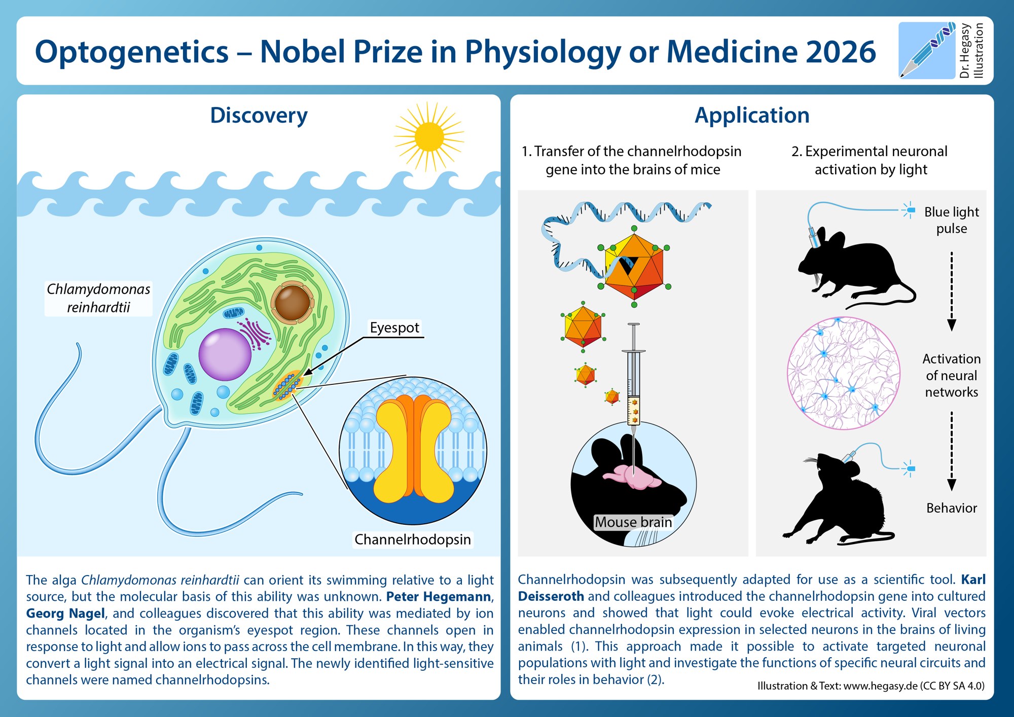
An explanatory illustration of the discovery of channelrhodopsin in Chlamydomonas reinhardtii and its use to activate nerve cells in mice with light. This is an independent illustration, not one issued by the Nobel committee. Illustration and text: Guido4 (Wikimedia Commons user), hegasy.de, own work, via Wikimedia Commons (CC BY-SA 4.0). Source page: https://commons.wikimedia.org/wiki/File:2026-Nobel-Prize-Channelrhodopsin-Hegasy_Illustration.png
The primary record is the Nobel Assembly at Karolinska Institutet's press release of 5 October 2026, "Nobel Prize in Physiology or Medicine 2026" (https://www.nobelprize.org/prizes/medicine/2026/press-release/). Additional details and Dr. Deisseroth's remarks are from Stanford University's announcement (https://news.stanford.edu/stories/2026/10/karl-deisseroth-nobel-prize-physiology-medicine). The Nobel Prize illustrations, licensed for non-commercial use only, are not reproduced here; the images above are Wikimedia Commons files.
- science
- medicine
- nobel-prize
- neuroscience
- optogenetics
- stanford